Abstract
Introduction. Pancreatic iron deposition is a common finding in thalassemia major, being detected in more than one third of patients undergoing their first T2* Magnetic Resonance Imaging scan (MRI) for this purpose. However, no longitudinal studies on pancreatic iron are available in literature.
Aim: The aim of this multicenter study was to evaluate the changes in pancreatic iron overload in TM patients enrolled in the Extension-Myocardial Iron Overload in Thalassemia (E-MIOT) Network who performed a baseline and a follow-up (FU) MRI scan at 18 months.
Methods. We considered 416 TM patients (37.77±10.46 years; 220 females) consecutively enrolled. Iron overload was quantified by the T2* technique. T2* measurements were performed over pancreatic head, body and tail and global value was the mean.
Results. Pancreatic iron overload (global pancreas T2*<26 ms) was detected in 367 (88.2%) patients. Of them, only 14 (3.8%) improved at the FU. Out of the 49 (11.8%) patients without baseline pancreatic iron overload, 15 (30.6%) showed pancreatic iron overload at the FU MRI.
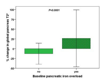
A significant inverse association was detected between % change in global pancreas T2*and baseline global pancreas T2* values (R=-0.369; P<0.0001). Patients with baseline pancreatic iron overload showed significantly higher % changes in global pancreas T2* values (see Figure).
Changes (%) in global pancreas T2* were not associated with baseline serum ferritin levels or MRI liver iron concentration (LIC) values but were inversely correlated with % changes in serum ferritin levels (R=-0.199; P<0.0001) and % changes in MRI LIC values (R=-0.255; P<0.0001). A significant positive association was found between % changes in global pancreas and global heart T2* values (R=0.133; P=0.007).
At baseline MRI, 169 patients showed an alteration of glucidic metabolism: 32 had impaired fasting glucose, 65 impaired glucose tolerance, and 72 diabetes mellitus. These patients showed significantly higher % changes in global pancreas T2* than patients with a normal glucidic metabolism (33.06±79.48% vs 11.93±59.47%; P=0.003).
Conclusions. Our data showed that it is difficult to remove the iron from the pancreas and higher improvements were detected in more heavily loaded patients, with alterations of glucidic metabolism. The reduction in pancreatic iron was paralleled by a decrease in hepatic and cardiac iron.
Pepe: Bayer S.p.A.: Other: no profit support; Chiesi Farmaceutici S.p.A: Other: no profit support. Maggio: Bluebird Bio: Membership on an entity's Board of Directors or advisory committees; Celgene Corp: Membership on an entity's Board of Directors or advisory committees; Novartis: Membership on an entity's Board of Directors or advisory committees.


This feature is available to Subscribers Only
Sign In or Create an Account Close Modal